Stanford's Aneurysm Center is a forerunner in the diagnosis and treatment of abdominal aortic aneurysms (AAAs). Performing an average of 100 AAA operations a year, our team of doctors, nurses, and other health care professionals strives to provide the most comprehensive, cutting edge care to this group of patients.
Our faculty also has extensive experience all FDA-approved devices for minimally invasive management of thoracic and abdominal aortic aneurysms as well as unique access to custom devices for particularly challenging clinical problems.
List of MD’s

Jason Lee, Vascular Surgeon

Ramford Ng, Interventional Cardiologist
Rajesh Dash, Cardiologist

Justin Lee, Cardiologist
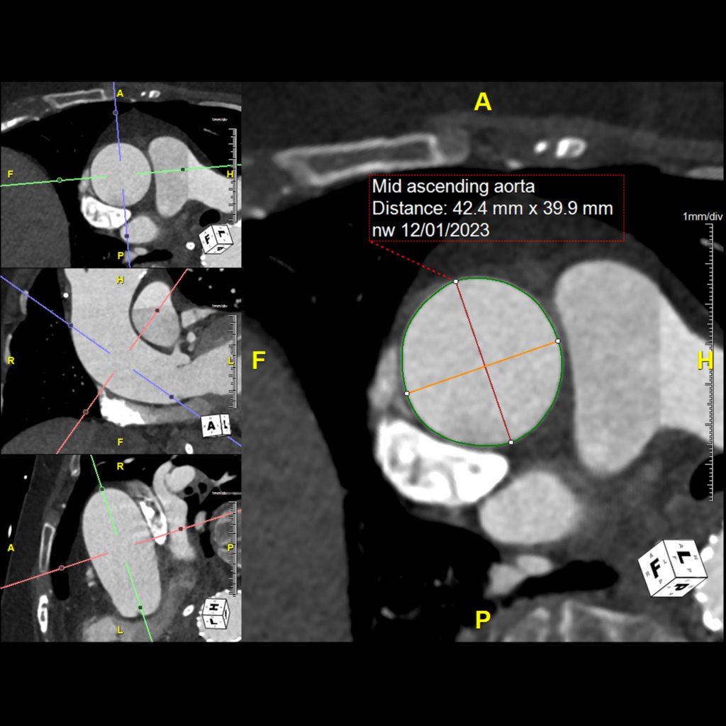
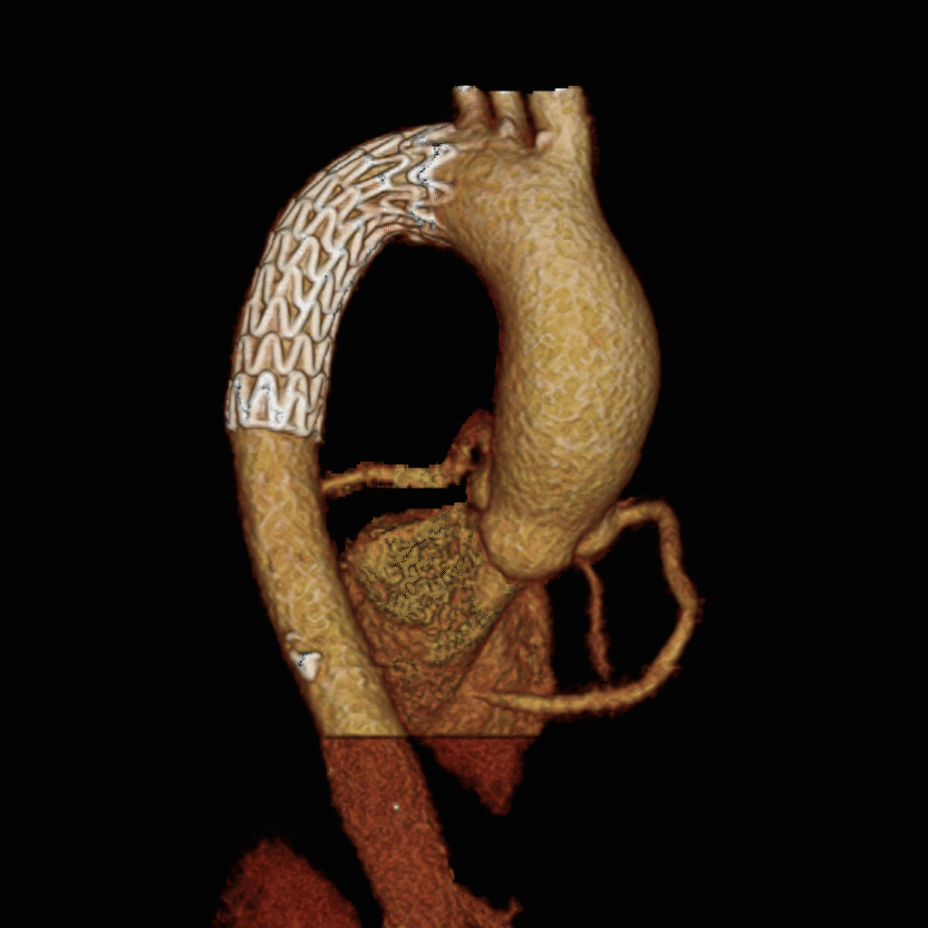
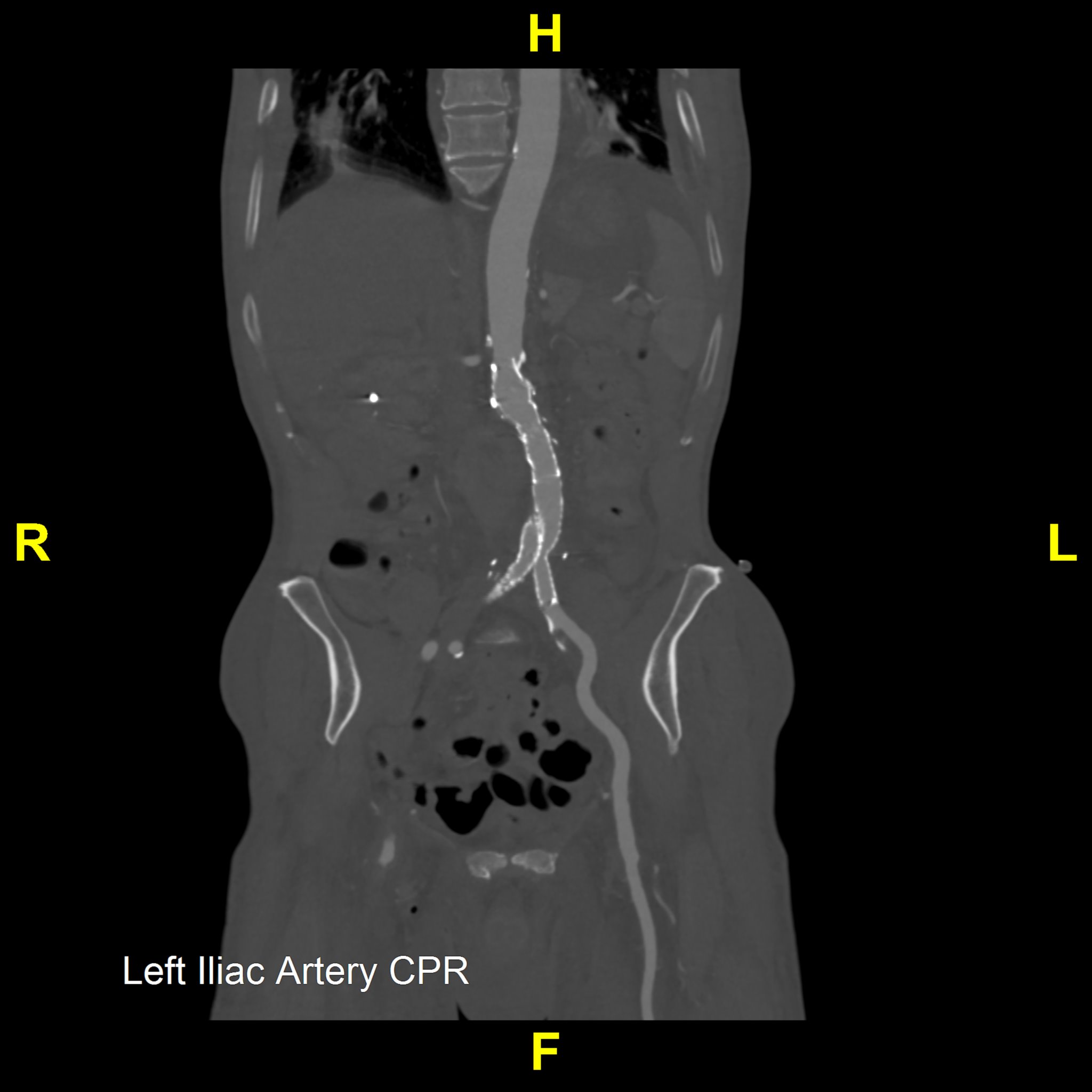
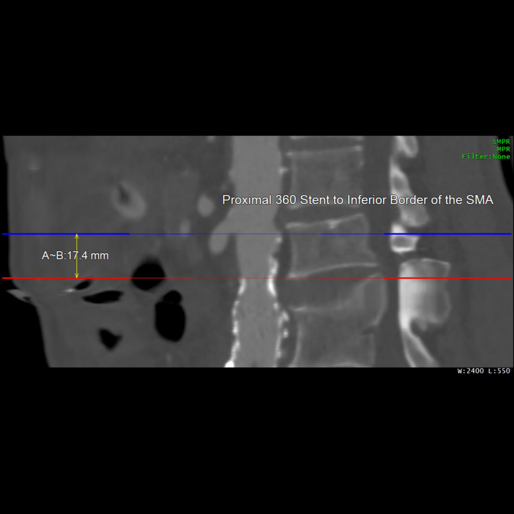
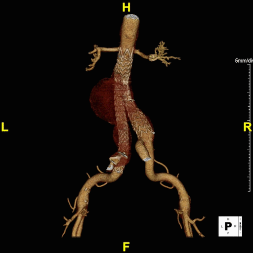
Our Post-Stent Service provides imaging for patients with stent placements in the abdominal aorta and common iliac arteries. The primary goal is to monitor changes in the maximum diameter of these vessels, ensuring early detection and management of post-stent issues. Using 3D imaging techniques identify critical aspects such as stent length, potential migration, and aortic diameter.
This protocol complements Routine Chest, Abdomen, and Pelvis imaging, offering an added layer of post-procedural monitoring. It allows quick viewing of major anatomy and focuses on identifying stent movement or aneurysm growth, ensuring comprehensive and effective monitoring.
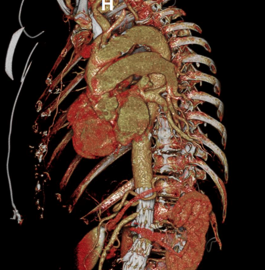
Our 3D Printing Service provides anatomical 3D printed models for surgical planning and education. 3D prints can be requested during the 3D imaging ordering process. To achieve optimal results we require high-resolution CT or MRI scans. Once the scan is obtained, we begin the process by segmenting the medical images to isolate specific anatomical structures. This segmented data is then converted into a 3D model and reviewed by the requestor.
Final models are produced using advanced 3D printing technologies such as Fused Deposition Modeling (FDM), Stereolithography (SLA), and PolyJet. We offer hand delivery for on-site requestors and mail delivery for those at other Stanford locations. Additionally, we are exploring the development of custom cutting guides to assist in surgical procedures.


Our Heart & Coronary Angiography service utilizes CT coronary angiography to assess the heart for hemodynamically significant calcific and soft plaques, as well as any anomalous anatomy, which may be useful for diagnosing and managing cardiovascular disease. We perform Curved Planar Reformation (CPR) on areas with minimal motion artifact, ensuring the clearest possible visualization of each coronary artery.
The service includes creating detailed CPRs for the Left Anterior Descending (LAD), Left Circumflex (LCX), Right Coronary Artery (RCA), and any bypass grafts, annotating native vessel names for understanding. We further enhance our evaluations with a 360-degree loop of each CPR providing an all-encompassing view of coronary anatomy.
For optimal clarity, our segmentation process selects the cardiac phase with the least motion, focusing solely on the heart to include muscle tissue, coronary arteries, and the proximal aorta. We remove extraneous structures only as necessary, preserving the integrity of the heart model.
Our service also offers a variety of captures, including lateral and AP views of the RCA, oblique views for LAD and LCX visualization, and superior views to demonstrate vessel origins, among others. These targeted views and visual reconstructed images allow for the detailed demonstration of vessels or pathology, assisting treatment strategies and improving patient outcomes in cardiovascular care.

