At Stanford Health Care, our electrophysiologists have an international reputation for treating people with atrial fibrillation and other arrhythmias, and they have invented many of today’s leading procedures. Our team specializes in ablation, surgical therapies, device management, and treatment of inherited conditions.


List of MD’s

Mohan Viswanathan, Cardiac Electrophysiologist

Paul Wang, Cardiac Electrophysiologist

Nitish Badhwar, Cardiac Electrophysiologist

Marco Perez, Cardiac Electrophysiologist
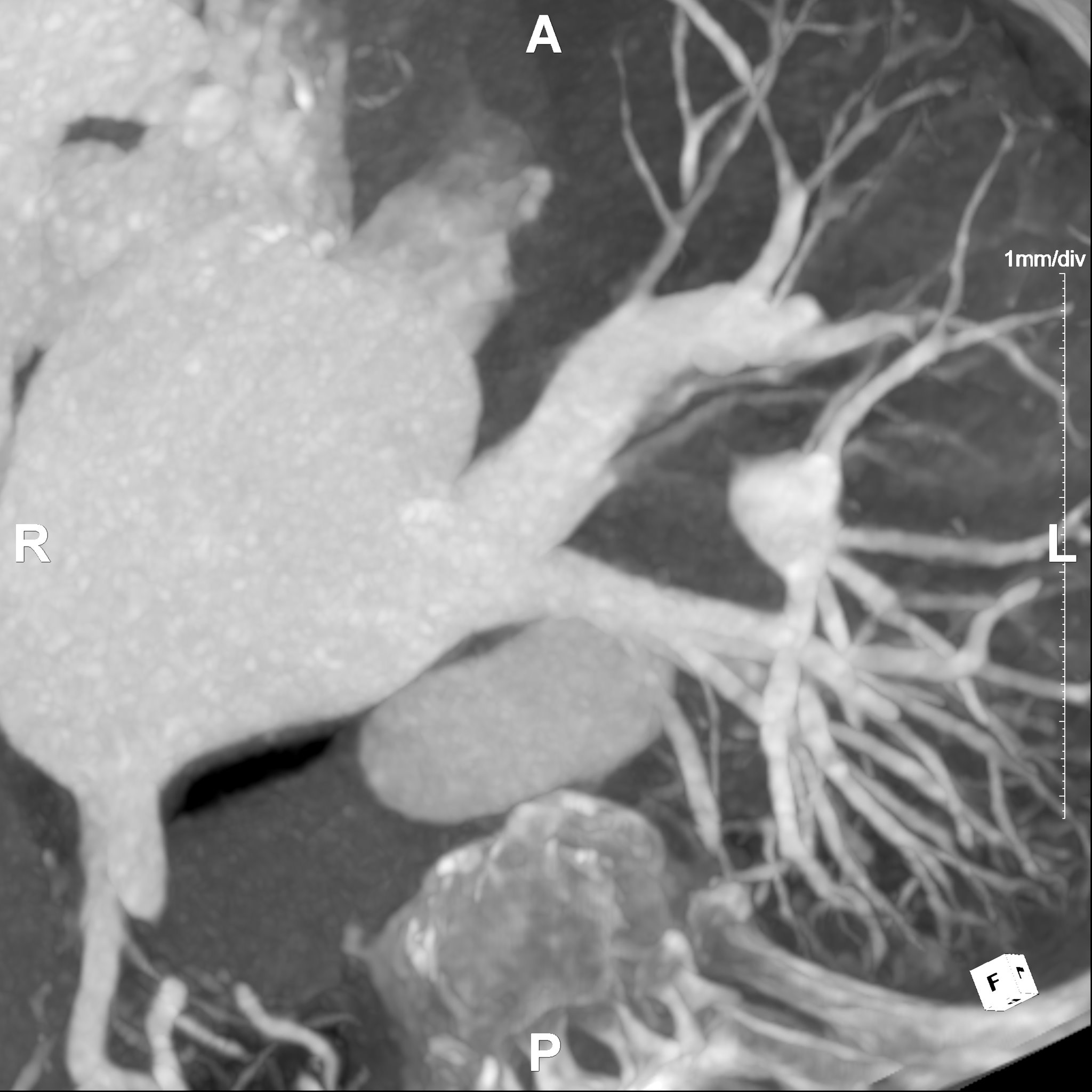
The Left Atrial Mapping service specializes in providing comprehensive left atrial imaging for the treatment of left atrial fibrillation (AFib) through ablation procedures. This service maps the pulmonary veins and their connections to the left atrium. This mapping can be used for pinpointing the origins of the erratic electrical signals that contribute to AFib. Maximum Intensity Projections (MIPs) enhance the visualization of the pulmonary veins by highlighting the brightest, most significant signals within the scan, corresponding to blood flow. This makes it easier to identify and target the specific areas responsible for AFib during the ablation process.
Additionally, the service provides measurements of orthogonal diameters and areas within the imaging data. These measurements offer an understanding of the pulmonary veins' dimensions and spatial relationships, providing insights for guiding the ablation procedure accurately. By analyzing these orthogonal measurements, specialists can assess the size and shape of the targeted areas, potentially ensuring a more focused and effective treatment. Post-ablation, the service continues its support by using these imaging techniques to assess for complications such as pulmonary vein stenosis.

Our 3D Printing Service provides anatomical 3D printed models for surgical planning and education. 3D prints can be requested during the 3D imaging ordering process. To achieve optimal results we require high-resolution CT or MRI scans. Once the scan is obtained, we begin the process by segmenting the medical images to isolate specific anatomical structures. This segmented data is then converted into a 3D model and reviewed by the requestor.
Final models are produced using advanced 3D printing technologies such as Fused Deposition Modeling (FDM), Stereolithography (SLA), and PolyJet. We offer hand delivery for on-site requestors and mail delivery for those at other Stanford locations. Additionally, we are exploring the development of custom cutting guides to assist in surgical procedures.


