Stanford Health Care’s Gynecologic Cancer Program combines compassion with innovation to treat all types of gynecologic cancers through surgery, chemotherapy, radiation, and medication. We also provide extensive supportive services, including genetic testing and counseling.
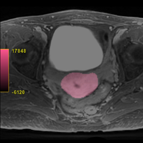
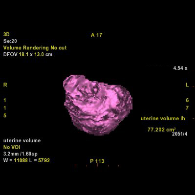
Our Uterine Fibroid Service offers an advanced 3D imaging solution designed to support the assessment and treatment planning for uterine fibroids. By accurately calculating the volume of the uterus and providing detailed mapping of the pelvic vasculature, this service may assist in the management of fibroids. Precise measurements of the uterus' volume are important in gauging the extent of fibroids present, which is essential for understanding the severity of the condition and planning appropriate interventions.
Additionally, this imaging may be useful for effective planning of uterine fibroid embolization (UFE), a minimally invasive procedure that aims to shrink fibroids by blocking their blood supply. This approach ensures that treatment is precisely targeted, maximizing efficiency and outcomes. UFE involves using imaging guidance to deliver small particles that obstruct the blood flow to fibroids, leading to their reduction.
Our service also provides follow-up measurements to monitor the response to various therapies, such as High-Intensity Focused Ultrasound (HIFU), UFE, or myomectomy, allowing for the adjustment of treatment plans based on the progress observed. With nearly 90 percent of women experiencing symptom relief from fibroids through such treatments, our Uterine Fibroid Service is committed to delivering targeted and effective solutions for fibroid management, enhancing patient care and outcomes.

Our Tumor Response Service, grounded in the Tumor Response Assessment Criteria (TRAC), plays a role in the fight against cancer by tracking tumor progression and response to treatments. TRAC reports, derived from advanced 3D imaging measurements, provide a detailed medical imaging perspective on tumor changes over time through longitudinal analysis. This method collects data points across the treatment timeline, offering a comprehensive view of the tumor's reaction to therapy and guiding healthcare professionals in making informed adjustments to treatment plans.
When a TRAC report is requested, our technologists first establish a baseline by measuring the dimensions of target and non-target tumors from initial scans. Follow-up scans, typically conducted three months into treatment, allow for repeated measurements and the accumulation of data analyzed according to specific tumor response criteria, tailored to the cancer type and treatment regimen. The analysis results, securely stored within our system, are presented in various formats such as tabulated summaries, stacked charts, and galleries of snapshots from DICOM images, enabling a multifaceted view of the patient's cancer journey.
Standardized tumor response criteria serve as the foundation for these evaluations, creating a consistent framework for assessing how tumors shrink, disappear, remain stable, or grow in response to therapy. This structured approach supports healthcare providers in evaluating treatment effectiveness, making informed decisions on continuing, modifying, or seeking alternative treatments based on the unique nature of the cancer, the treatment strategy, and the evolving landscape of cancer therapies. Our service, through TRAC, empowers clinicians with precise insights to optimize patient outcomes in the complex fight against cancer.

