We use interventional cardiology to diagnose many heart and vascular problems. Interventional cardiology also helps us avoid surgery with treatments like angioplasty and valve repair or replacement. Our doctors invented many technologies used today and they continue to innovate the field to advance your care options.

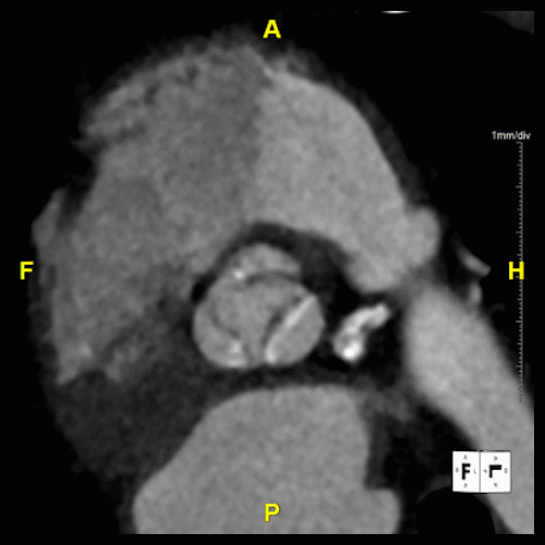
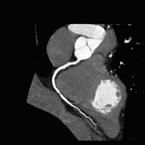


Our Heart & Coronary Angiography service utilizes CT coronary angiography to assess the heart for hemodynamically significant calcific and soft plaques, as well as any anomalous anatomy, which may be useful for diagnosing and managing cardiovascular disease. We perform Curved Planar Reformation (CPR) on areas with minimal motion artifact, ensuring the clearest possible visualization of each coronary artery.
The service includes creating detailed CPRs for the Left Anterior Descending (LAD), Left Circumflex (LCX), Right Coronary Artery (RCA), and any bypass grafts, annotating native vessel names for understanding. We further enhance our evaluations with a 360-degree loop of each CPR providing an all-encompassing view of coronary anatomy.
For optimal clarity, our segmentation process selects the cardiac phase with the least motion, focusing solely on the heart to include muscle tissue, coronary arteries, and the proximal aorta. We remove extraneous structures only as necessary, preserving the integrity of the heart model.
Our service also offers a variety of captures, including lateral and AP views of the RCA, oblique views for LAD and LCX visualization, and superior views to demonstrate vessel origins, among others. These targeted views and visual reconstructed images allow for the detailed demonstration of vessels or pathology, assisting treatment strategies and improving patient outcomes in cardiovascular care.


Our service for Transcatheter Aortic Valve Replacement (TAVR) utilizes 3D imaging and measurements to enhance procedural planning. These images may be useful for mapping the intricate landscape of cardiac structures and vasculature, ensuring each transcatheter heart valve (THV) procedure is customized to the patient's unique anatomy.
Using 3D imaging in the TAVR planning process allows for the evaluation of aortic root dimensions, annulus size, and the morphology of the aortic valve complex. It provides insights into the spatial relationships between the aortic valve and surrounding coronary arteries, highlighting potential challenges and guiding the selection of the appropriate valve size and optimal implantation approach.
Additionally, CT angiography assesses the iliofemoral arteries, determining their suitability for catheter access by identifying any calcifications, occlusions, or tortuosities that could impact the procedure.


