The Nephrology Clinic specializes in the diagnosis and treatment of kidney disorders.

List of MD’s

Alan Thong, Urologic Surgeon

Eila Skinner, Urologic Oncologist

Craig Comiter, Urologist

Timothy Chang, Urologist
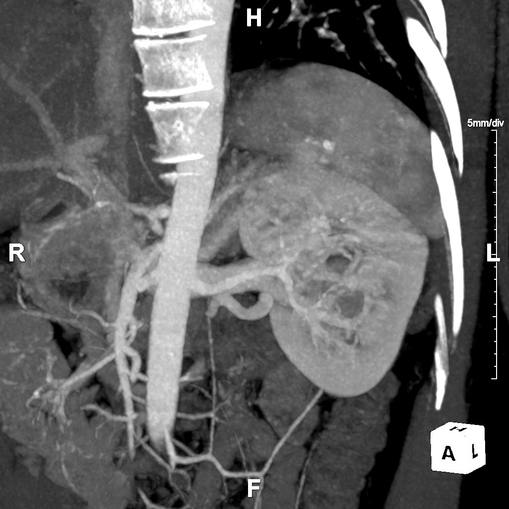
Our Routine Chest Abdomen Pelvis Service provides 3D reconstructions focusing on a thorough evaluation of the vascular system in the chest, abdomen, or pelvis. This protocol is intended for use on cases where the referring physician does not have a specialized protocol. If the patient's history is unclear, this is the default protocol for all chest, abdomen, and pelvis angiograms, or any combination thereof. Our aim for this protocol is to allow quick viewing of major anatomy and then focus only on areas of concern.
Imaging methods like Maximum Intensity Projections (MIPs) and Curved Planar Reformation (CPRs) enhance the clarity of vascular structures. MIPs are effective for visualizing blood vessels by projecting the highest attenuation values onto a 2D image, while CPRs offer detailed views along the natural curves of the vessels, aiding in the detection of abnormalities and enhancing diagnostic accuracy.


Our Urology Service utilizes MR Urogram with advanced 3D imaging technology to provide three-dimensional reconstructions of the urinary tract. By employing MRI, we are able to harness the power of T2-weighted imaging, which is particularly effective for the clear visualization of fluid-filled structures within the urinary system. This technique ensures that areas such as the bladder, ureters, and kidneys are depicted with high clarity, aiding in the comprehensive assessment of these critical structures.
To further enhance the diagnostic capability of our imaging, we may also include post-contrast sequences. These sequences delineate the urothelium, the layer of tissue lining the urinary tract, and for the detection of any lesions or abnormalities. This dual approach, combining T2-weighted and post-contrast imaging, may provide a thorough evaluation of the urinary tract's health, highlighting both the normal anatomical features and any potential issues that may require closer examination or intervention.
Through our Urology Service, we assist healthcare professionals by providing images that may support the diagnosis and management of urological conditions, facilitating targeted treatments and improved patient outcomes.

Our Kidney Stone Service utilizes dual-energy CT scans to assist in treatment planning by distinguishing between different types of kidney stones based on density differences observed under varying kilovoltage (kV) settings. This technique provides detailed imaging to accurately characterize calcifications and determine the most appropriate treatment approach.
By analyzing density ratios of lesions, particularly those exceeding a 1.13 ratio, our service identifies stones too dense for lithotripsy therapy, indicating the need for surgical intervention. This differentiation ensures patients receive the treatment best suited to their specific condition.
