Stanford Department of Neurosurgery is home to 60 leading neurosurgeons and research faculty. Our neurosurgeons perform over 4,000 neurosurgical operations covering the full spectrum of neurological conditions every year. We diagnose, treat, and cure neurological conditions with the precision necessary to restore patients to their lives.


List of MD’s
Neil Schwartz, Stroke & Vascular Neurologist
Vivek Buch, Neurosurgeon

Jaimie Henderson, Functional Neurosurgeon
Gary Steinberg, Cerebrovascular Neurosurgeon
Our Functional MRI (fMRI) service uses advanced technology to create detailed maps showing how different parts of the brain function. This method tracks changes in blood flow and oxygen levels in the brain while a person performs specific tasks or rests. It helps identify active areas of the brain responsible for functions like movement, speech, hearing, and seeing. This can be especially important for understanding how brain disorders such as tumors or strokes might affect these functions.
The collected data is presented in detailed visualizations of brain activity and connectivity. We offer various imaging views, such as 3D color maps and streamline images, which show how different brain regions connect and communicate. These images may be important for planning surgeries to remove brain lesions safely without damaging these essential functions.


The Bony Head service specializes in 3D imaging for assessing craniofacial abnormalities. This service focuses on evaluating conditions such as the premature closure of skull sutures, abnormal skull shapes, and growth abnormalities affecting the craniofacial region.
Our service offers a suite of 3D imaging options, including cut plane visual reconstruction (VR) views, rotational VR movies, and cut plane VR movies, providing a dynamic and in-depth analysis of craniofacial anatomy. These technologies offer cross-sectional views and 360-degree tours around the cranial structure, promoting the detection and assessment of abnormalities in skull shape or suture closure.
These 3D imaging techniques support a thorough evaluation and understanding of various craniofacial abnormalities, aiding in accurate diagnosis and the planning of appropriate interventions.


Our Brain Fiber Tracking service provides imaging of the brain's wiring by using an imaging technique called 3D Diffusion Tensor Imaging (DTI). This service is designed to map out the neural pathways of the brain, providing a visualization of how different regions connect and communicate.
This service is performed by combining DTI with standard imaging from CT and MRI scans. The alignment of these images ensures that we can accurately position and map the neural pathways, capturing the movement of water molecules along the brain's white matter tracts. These tracts are important for transmitting signals throughout the brain and are involved in everything from motor control to sensory perception. Our technique enhances these images with color coding and streamlined visuals, making the complex structure of the brain's connections more accessible and understandable.
The benefits of undergoing our Brain Fiber Tracking service are potentially significant, especially for clinical and research purposes. Functional MRI (fMRI), which is used in conjunction with DTI, allows us to see active brain areas by monitoring changes in blood flow during specific tasks. This capability is useful for precise mapping of active neural pathways, aiding in pre-surgical planning and the assessment of conditions like brain tumors or epilepsy. By identifying and preserving critical neural functions, this service can greatly improve surgical outcomes and contribute to more effective management of neurological conditions.


Our Focused Ultrasound Therapy Service offers imaging to aid in the treatment of essential tremor (ET), a condition characterized by involuntary shaking. Using Magnetic Resonance Imaging (MRI) technology, we create detailed 3D images of the brain, concentrating on areas critical for movement control.
These imaging outputs highlight the brain's pathways, helping doctors understand and target specific structures. This is important because essential tremor primarily affects brain regions responsible for motor functions, such as the thalamus. Accurate imaging ensures that focused ultrasound therapy can precisely target these areas, disrupting abnormal brain activity that causes tremors. This non-invasive treatment uses sound waves to reduce tremors without the need for surgical skull openings, offering a safer alternative to traditional surgery.
By delivering clearer views of brain pathways, our imaging techniques aims to enhance the accuracy and effectiveness of focused ultrasound therapy. This precision leads can lead to better control of tremors and improved patient outcomes, significantly enhancing the quality of life for those affected by ET.


Our Neurosurgical Guidance service leverages augmented reality (AR) to transform surgical planning. Surgeons can visualize and manipulate 3D segmentations of critical structures on interactive touch screens and VR headsets, providing a comprehensive understanding of the patient's anatomy before and during surgery.
This service is adept at handling complex neurosurgical cases, such as arteriovenous malformations (AVMs), aneurysms, cavernous malformations, and tumor pathologies. The integration of imaging with augmented reality neurosurgery navigation systems assists with the planning and execution of surgical interventions, making the intricate anatomy and pathology spatially relevant and easily accessible during procedures.
Extended reality technologies may offer significant advantages in neurosurgery. Surgeons can simulate different surgical scenarios to identify the most effective approach. This preoperative visualization can help plan the optimal surgical route to minimize invasiveness and avoid critical areas, thereby enhancing patient safety and surgical outcomes.

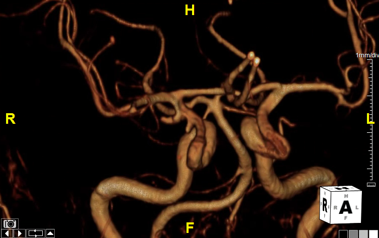
3D imaging for head and neck angiograms is a diagnostic tool that significantly enhances the detection and assessment of vascular conditions such as cerebral aneurysms, and stenosis, with a particular focus on the Circle of Willis. This imaging process allows providers to see the presence of calcium within aneurysms, the specific configuration of aneurysm necks, and the origins of vital vessels. Understanding these details is crucial for determining the most appropriate treatment pathway for patients.
These renderings also reveal stenosis or dissections in carotid or vertebral arteries, providing a view that may aid in diagnosis and treatment planning. These images are available within 45 minutes after CT acquisition, ensures timely decision-making.
This service creates many series of images. This includes axial, coronal, and sagittal maximum intensity projections (MIPs), as well as curved planar reconstructions (CPRs) of the left and right carotid arteries. Additionally, volumetric rendering (VR) of the Circle of Willis offers insights into the vascular architecture, aiding in the comprehensive evaluation of these structures.


