Serving over 2.6 million people, Stanford Medicine is the only Level 1 Adult and Level 1 Pediatric Trauma Center verified by the American College of Surgeons (ASC) on the peninsula of the San Francisco Bay Area. We provide specialized care to over 3700 patients per year and handle 20-25 consults daily. We offer exceptional 24-hours-a-day onsite trauma specialists in a state-of-the art facility, fully equipped to handle any medical or surgical emergency.

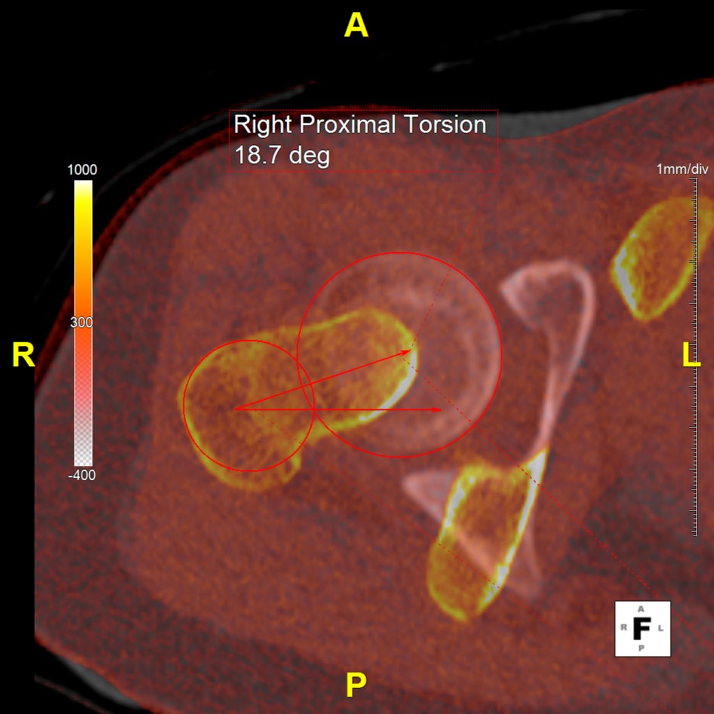
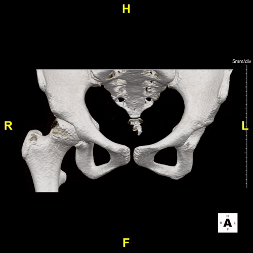
Our Bony Extremities Service specializes in 3D imaging of the lower and upper extremities, transforming traditional 2D CT or MRI scans into three-dimensional reconstructions. These images are can be useful for visualizing the complex bony structures of the leg, ankle, foot, shoulder, arm, elbow, wrist, and hand, enhancing diagnostic accuracy and surgical planning.
For lower extremity imaging, our service provides detailed views of the leg, ankle, and foot, useful for assessing fractures and planning surgeries. For upper extremities, we offer imaging of the shoulder, arm, elbow, wrist, and hand, aiding in the diagnosis and treatment of various conditions.
Additionally, our service is valuable for planning Anterior Cruciate Ligament (ACL) surgeries. We create 3D reconstructions of the knee joint, offering clear views of bones, existing grafts, and the joint's overall geometry to support precise surgical planning.


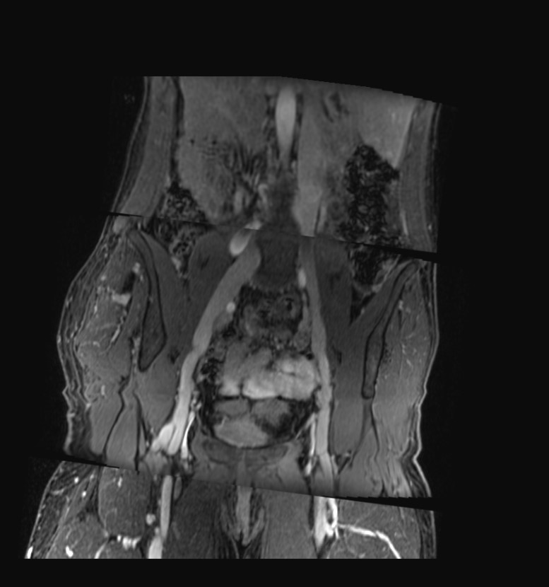
Our Venogram Service utilizes CT venography to offer a comprehensive view of the venous system, including the abdomen, pelvis, and lower extremities. Employing advanced 3D imaging techniques such as Maximum Intensity Projection (MIP) batches and Curved Planar Reformation (CPR), we aim to enhance the visualization of vascular structures throughout these regions. This approach may be useful for ensuring detailed imaging from the thoracic veins down to the veins of the lower limbs, providing a thorough examination of the venous system.
In addition to CT venography, our service incorporates MR venography, which is specifically valuable for displaying the venous system in the abdomen, pelvis, and lower extremities. This modality is particularly adept at assessing various venous conditions, including stenosis, the presence and condition of stenting, post-thrombotic changes, varicose alterations, and evaluating the saphenous vein for bypass suitability.
This dual approach may assist healthcare professionals to make informed decisions regarding treatment options, based on a comprehensive understanding of the patient's venous health, thereby enhancing patient care and outcomes in venous disease management.


3D imaging for head and neck angiograms is a diagnostic tool that significantly enhances the detection and assessment of vascular conditions such as cerebral aneurysms, and stenosis, with a particular focus on the Circle of Willis. This imaging process allows providers to see the presence of calcium within aneurysms, the specific configuration of aneurysm necks, and the origins of vital vessels. Understanding these details is crucial for determining the most appropriate treatment pathway for patients.
These renderings also reveal stenosis or dissections in carotid or vertebral arteries, providing a view that may aid in diagnosis and treatment planning. These images are available within 45 minutes after CT acquisition, ensures timely decision-making.
This service creates many series of images. This includes axial, coronal, and sagittal maximum intensity projections (MIPs), as well as curved planar reconstructions (CPRs) of the left and right carotid arteries. Additionally, volumetric rendering (VR) of the Circle of Willis offers insights into the vascular architecture, aiding in the comprehensive evaluation of these structures.


