We provide the highest quality clinical care for patients dealing with vascular disease. Our interdisciplinary team excels in designing innovative solutions to challenging vascular problems, whether it's medical or surgical management, aneurysms, or aortic dissections.


List of MD’s

Jason Lee, Vascular Lee

John Higgins, Pathologist

Andrew Chu, Emergency Medicine Doctor

Al'ai Alvarez, Emergency Medicine Doctor
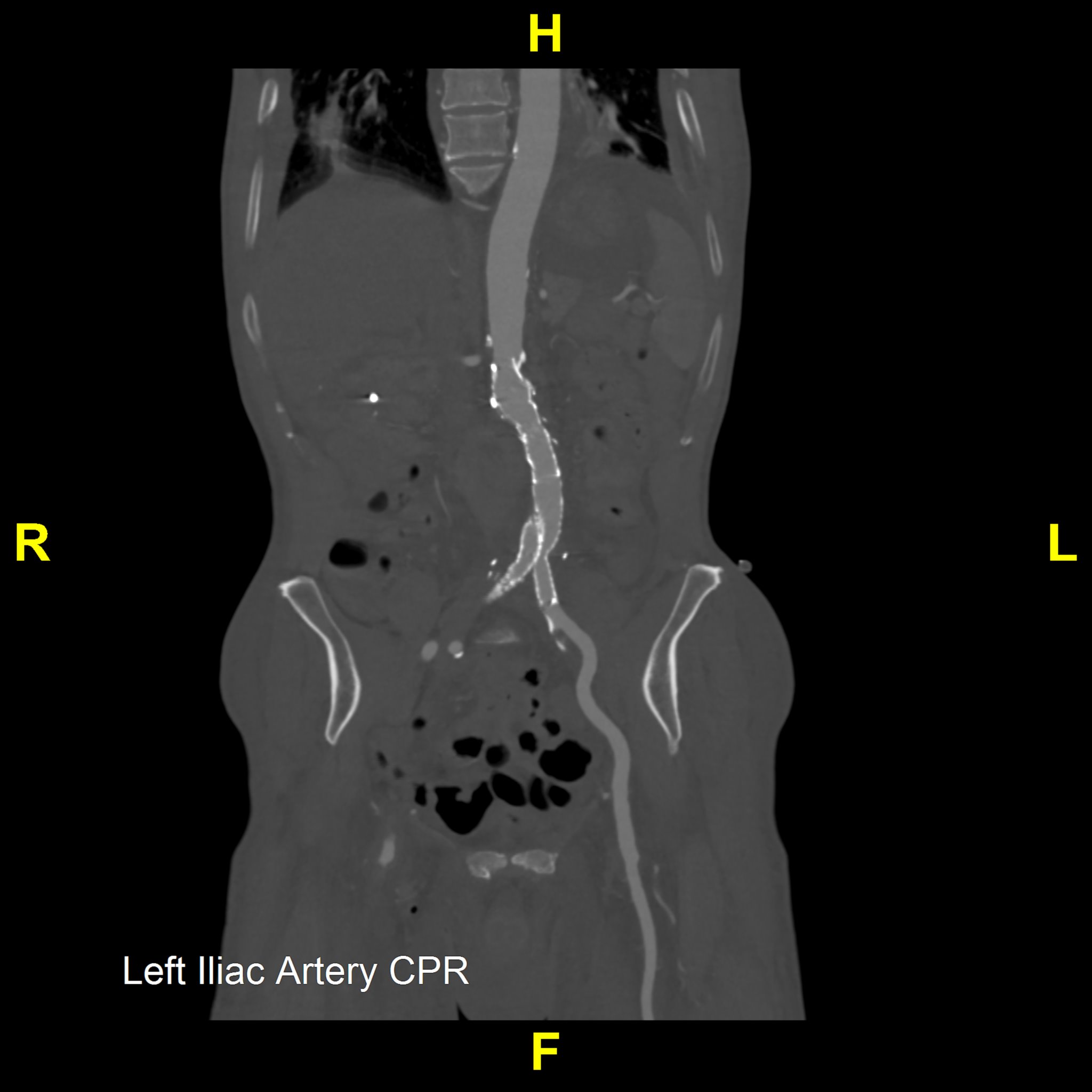
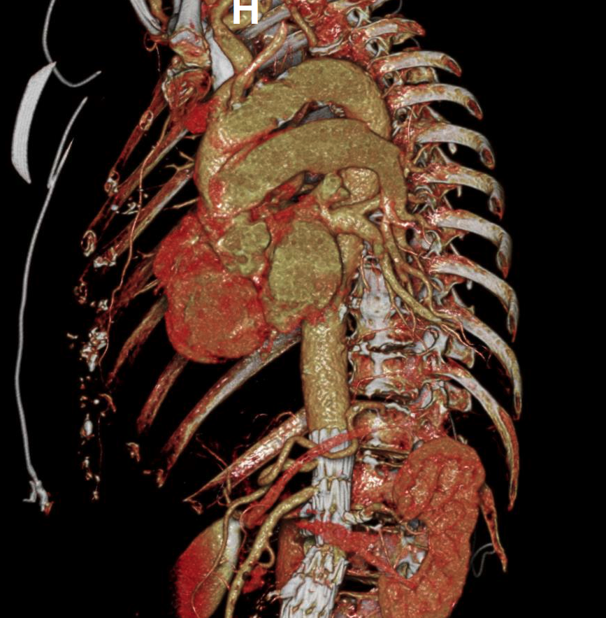
Our Venogram Service utilizes CT venography to offer a comprehensive view of the venous system, including the abdomen, pelvis, and lower extremities. Employing advanced 3D imaging techniques such as Maximum Intensity Projection (MIP) batches and Curved Planar Reformation (CPR), we aim to enhance the visualization of vascular structures throughout these regions. This approach may be useful for ensuring detailed imaging from the thoracic veins down to the veins of the lower limbs, providing a thorough examination of the venous system.
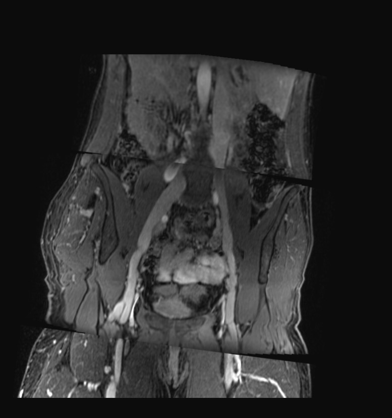
In addition to CT venography, our service incorporates MR venography, which is specifically valuable for displaying the venous system in the abdomen, pelvis, and lower extremities. This modality is particularly adept at assessing various venous conditions, including stenosis, the presence and condition of stenting, post-thrombotic changes, varicose alterations, and evaluating the saphenous vein for bypass suitability.
This dual approach may assist healthcare professionals to make informed decisions regarding treatment options, based on a comprehensive understanding of the patient's venous health, thereby enhancing patient care and outcomes in venous disease management.


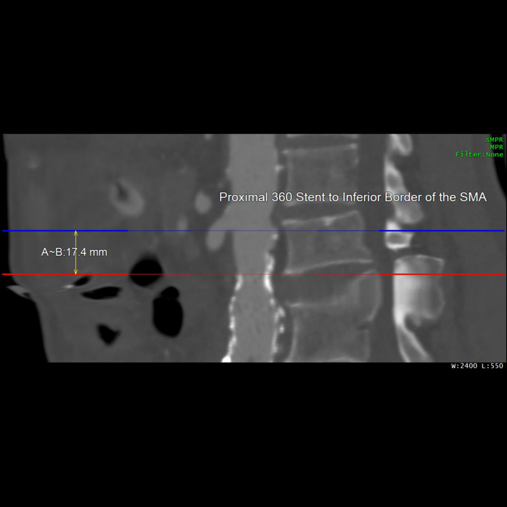
Our Post-Stent Service provides imaging for patients with stent placements in the abdominal aorta and common iliac arteries. The primary goal is to monitor changes in the maximum diameter of these vessels, ensuring early detection and management of post-stent issues. Using 3D imaging techniques identify critical aspects such as stent length, potential migration, and aortic diameter.
This protocol complements Routine Chest, Abdomen, and Pelvis imaging, offering an added layer of post-procedural monitoring. It allows quick viewing of major anatomy and focuses on identifying stent movement or aneurysm growth, ensuring comprehensive and effective monitoring.
Our Left Ventricular Assist Device (LVAD) Service uses CT coronary angiography to evaluate the placement of LVAD and Right Ventricular Assist Device (RVAD) in patients. LVADs and RVADs are mechanical pumps that support heart function in patients with severe heart failure, often serving as a temporary solution while awaiting heart transplantation. This imaging technique provides targeted views of the heart and surrounding blood vessels, ensuring these devices are correctly positioned. Proper placement is crucial for the functionality of these devices, minimizing complications, and improving overall outcomes in advanced cardiac care.


Our Lower Extremity Angiography service is tailored to detect occlusions and collaterals in the aortoiliac, femoral, popliteal, and lower leg vessels. We offer targeted imaging of the lower leg vasculature for pre-surgical planning, particularly for fibular free flap procedures, supporting the diagnosis and treatment of vascular conditions. These images provide clear visualizations of arterial pathways, including any abnormal courses or anatomical variations that could cause vascular entrapment or compromise.


